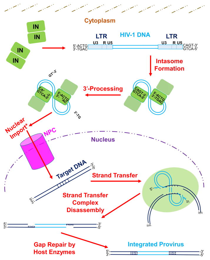
Figure 1. The mechanism of retroviral integration.
The integration process begins in the host cell with formation of the intasome, which consists of multimerized IN bound to the viral LTR ends. The LTR is composed of U3, R, and U5 sections, each of which contains unique elements responsible for mediating viral gene transcription (Pereira et al., 2000). While within the cytoplasm, IN cleaves nucleotides (specifically two nucleotides for the depicted HIV-1 integration pathway) from each 3′ end of the vDNA adjacent to invariant 5′-CA-3′ dinucleotides during 3′-processing. Completion of 3′-processing leaves a recessed and chemically reactive hydroxyl group at each vDNA 3′ end. Lentiviral PICs are actively imported into the cell nucleus (denoted by an asterisk in figure), while γ-retroviruses can only enter the nucleus after dissolution of the nuclear membrane during cell division [reviewed in: (Matreyek and Engelman, 2013)]. Within the nucleus, IN binds tDNA and utilizes the reactive vDNA 3′-hydroxyl groups to simultaneously cleave the tDNA phosphodiester backbone and insert the vDNA molecule, through the process of strand transfer. IN subunits cleave the top and bottom tDNA strands in a staggered fashion. The length of stagger differs among retroviruses; HIV-1 IN cleaves tDNA with the depicted 5-bp stagger. After disassembly of the strand transfer complex, host cell enzymes repair the DNA recombination intermediate to yield the integrated provirus flanked by a host DNA TSD. See main text for descriptions of the roles that other cellular factors play in the process of retroviral DNA integration. Please note that a color version of Figure 1 is available online.

