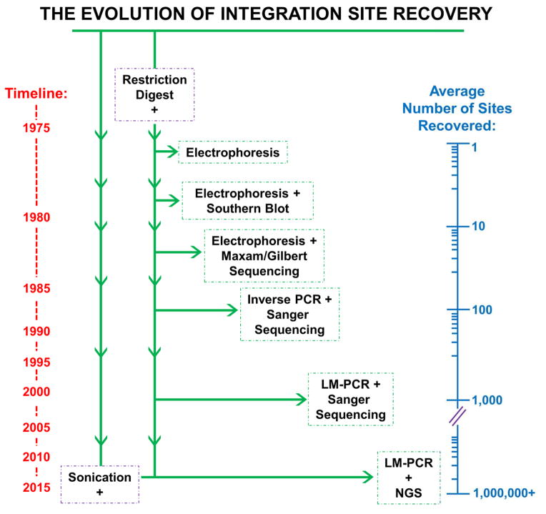
Figure 2. The evolution of techniques used to determine retroviral integration sites.
Integration site recovery techniques are listed in chronological order from top to bottom, corresponding to a general timeline shown at the left of the figure. The average numbers of integration sites recovered in studies employing these techniques, which exponentially increased over time, are illustrated on a logarithmic axis at the right side of the figure. The initial step of host cell gDNA fragmentation by restriction endonuclease digestion is shared by all of the listed techniques, though sonication has recently become a desirable method of shearing. Please note that a color version of this figure is available online.

