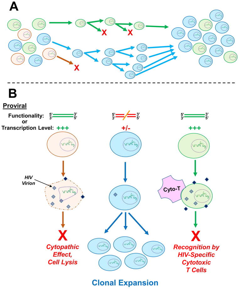
Figure 4. Clonal expansion of provirus-harboring cells.
A) General schematic of cellular clonal expansion. From an initially diverse population of cells, certain clones can exhibit relatively increased viability or growth rate, thus leading to persistence and ultimate enrichment. In this example the orange cell clone is dividing slowly or not at all, while the green cell clone exhibits an intermediate rate of expansion, and the blue cell clone expands most quickly. Thus in the final cell population, the blue clone predominates. B) Host cells harboring a particular integration site(s) can also become enriched over time within infected patients. The fact that clonally expanded, provirus-harboring cells can persist in the face of suppressive ART suggests that additional factors beyond integration site-mediated stimulation of cell growth rate contribute to clonal persistence. Two suggestions have been that the resident proviruses within the expanded cell clones are 1) nonfunctional due to non-tolerable insertions or deletions or 2) are molecularly intact but are expressed at a relatively low level (or not at all). In either case, the clones would escape viral replication-associated cytopathic effects and cell lysis (orange clone), as well as host cell recognition and purging by HIV-specific cytotoxic T-cells (green clone), leading to the enrichment of particular integration sites over time (blue clone). Neither of these possibilities, though, has thus far been definitively proven. A color version of this figure is available online.

