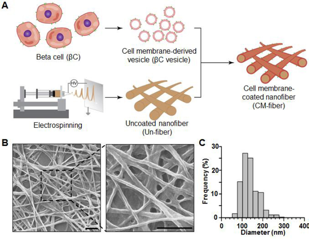
Fig. 1.
Preparation and characterization of cell membrane-coated nanofibers (CM-fibers). (A) A schematic illustration showing the preparation of CM-fibers. The process can be divided into three steps: deriving membrane vesicles from beta cells (βC vesicles), fabricating uncoated nanofibers (Un-fibers) via an electrospinning method, and fusing the βC vesicles onto the surface of the Un-fibers. (B) Representative SEM images depicting the fibrous morphology of the resulting CM-fibers (scale bar, 1 µm). (C) Size and size distribution of the CM-fibers.

