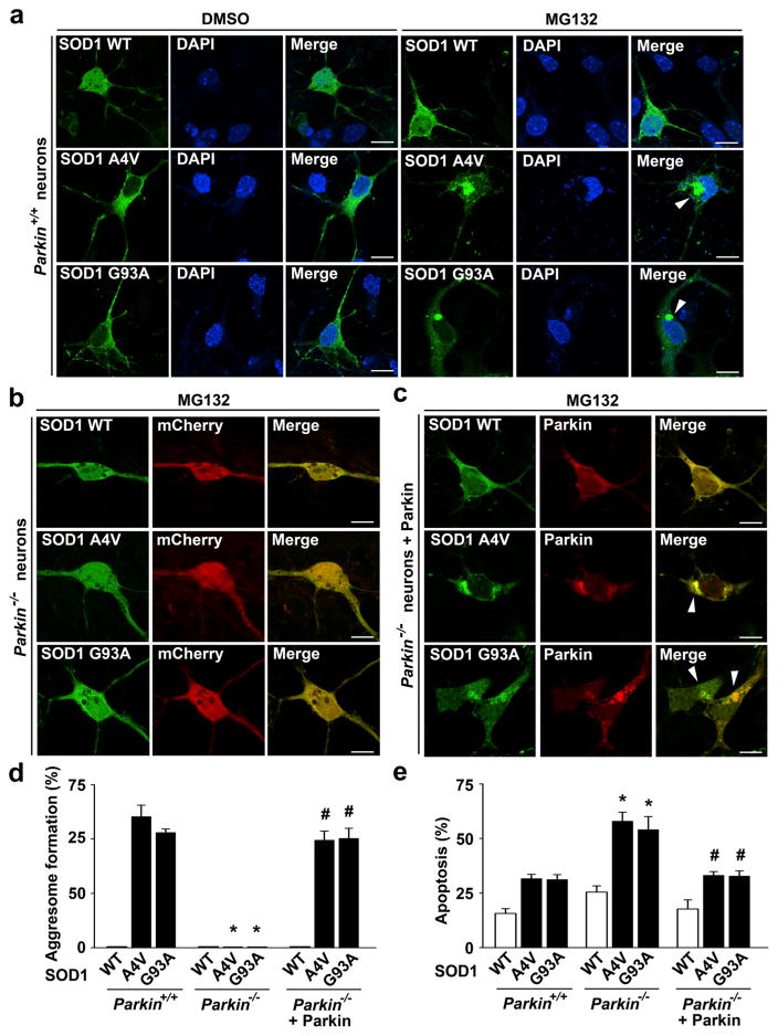
Fig. 10.
Targeted parkin deletion abolishes mutant SOD1 aggresome formation and enhances neuronal vulnerability to mutant SOD1-induced toxicity. a–c Parkin+/+ and parkin−/− cortical neurons transfected with indicated GFP-tagged SOD1 WT or mutant and mCherry or mCherry-parkin were treated with the vehicle DMSO or 5 μM MG132 for 24 h as indicated and then imaged by fluorescence confocal microscopy. Merged channel in (a) shows GFP-SOD1 (green) and DAPI-stained nuclei (blue). Scale bar=10 μm. d Aggresome formation was quantified and expressed as the percentage of SOD1-transfected cells containing SOD1-positive aggresomes. *P<0.05 versus the corresponding parkin+/+ control; #P<0.05 versus the corresponding mCherry-transfected parkin−/− neurons, two-way ANOVA with Tukey’s post hoc test. e Parkin+/+ and parkin−/− cortical neurons transfected with indicated GFP-tagged SOD1 WT or mutant and mCherry or mCherry-parkin were treated with 5 μM MG132 for 24 h, and nuclear integrity was assessed by DAPI staining. Apoptosis is expressed as the percentage of SOD1-transfected cells with apoptotic nuclear morphology. Data represent mean ± SEM from three independent experiments. *P<0.05 versus the corresponding control level in parkin+/+ neurons; #P<0.05 versus the corresponding level in parkin−/− neurons, two-way ANOVA with Tukey’s post hoc test

