Fig. 8.
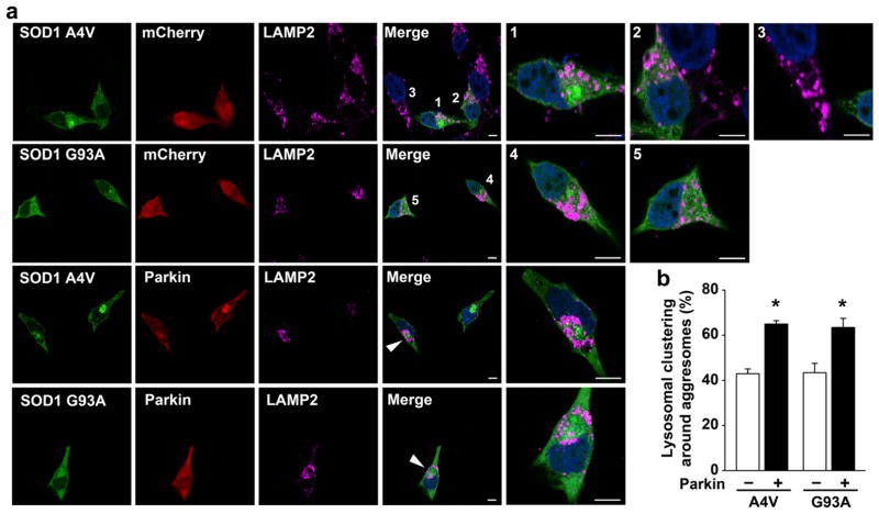
Parkin promotes lysosome clustering around mutant SOD1 aggresomes. a SH-SY5Y cells transfected with mCherry or mCherry-parkin and GFP-SOD1 A4V or G93A were treated with 5 μM lactacystin for 16 h and then processed for immunofluorescence confocal microscopic analysis with anti-LAMP2 antibody to visualize lysosome positioning. Merged channel includes GFP-SOD1 (green), LAMP2-positive lysosomes (purple), and DAPI-stained nuclei (blue). The numbers or arrowheads indicate the cells shown in the enlarged view. Scale bar =10 μm. b Quantification of the percentage of aggresomes that have lysosomal clustering around them. *P<0.05 versus the corresponding mCherry control cells lacking exogenous parkin, unpaired two-tailed Student’s t test

