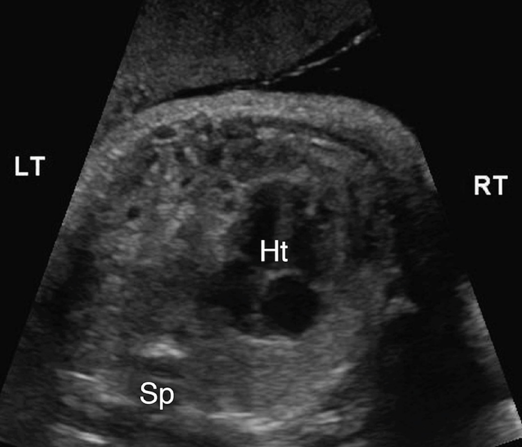
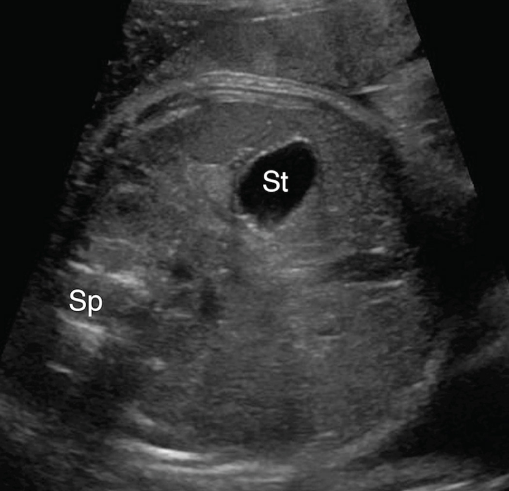
Figure 1. 1A & 1B. Intra-abdominal stomach position.
A. Transaxial gray-scale sonographic image of the chest in a 31.4-week fetus. Bowel loops herniated into the left chest displace the heart (Ht) to the right. The stomach is not seen within the chest.
B. Evaluation of the fetal abdomen demonstrated normal intra-abdominal location of the stomach (St). Spine (Sp).


