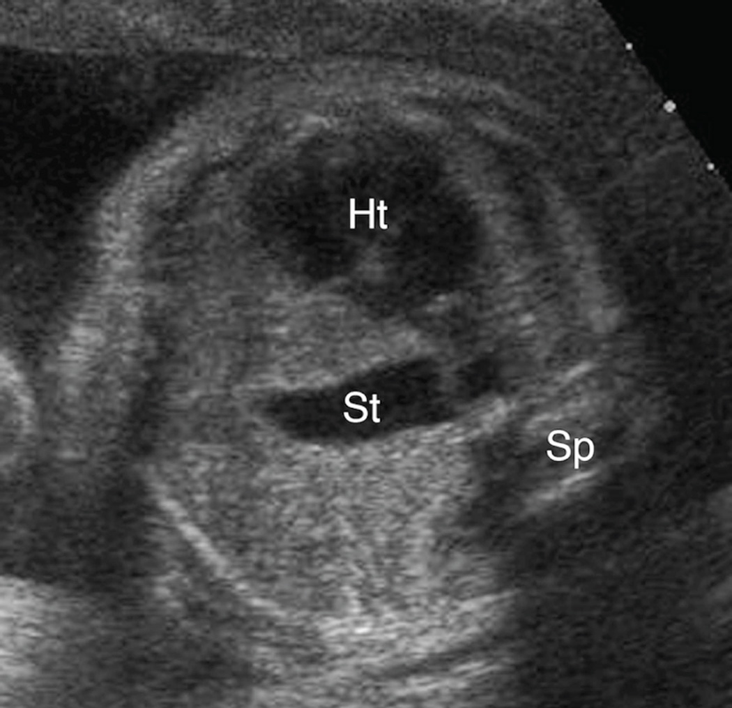
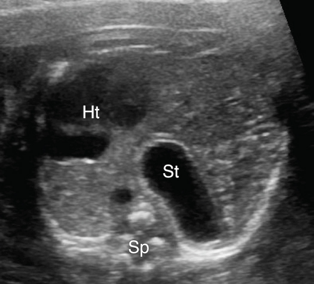
Figure 3. A & 3B. Spectrum of mid-to-posterior left chest stomach position.
A. Transaxial gray-scale sonographic image of the chest in a 32-week-old fetus. The obliquely oriented stomach (St) contacts neither the anterior nor posterior chest walls and remains entirely within the mid portion of the left chest. Heart (Ht), spine (Sp).
B. Transxial gray-scale sonographic image of the chest in a 20.7-week fetus. Herniated stomach (St) contacts the posterior wall of the left chest. Heart (Ht), spine (Sp).


