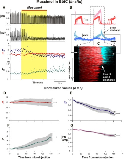
Figure 14.
Disturbances of respiratory pattern including elimination of post-inspiratory activity by bilateral microinjection of muscimol (10 µm) in the BötC of juvenile rat perfused brainstem–spinal cord in situ. A, Representative example of augmented ∫PN inspiratory discharge frequency and pronounced reduction of cVN integrated (∫cVN) activity amplitude due to suppression of post-I activity. Overlaid traces in B and time-series raster plot in C of consecutive respiratory cycles aligned to inspiratory onset (indicated by vertical dotted line in B) illustrate perturbations of cVN post-I discharge during BötC muscimol microinjection in another experiment. B, Integrated PN (red traces) and cVN (blue traces) activity are shown before (lighter red and lighter blue traces, respectively) and after (dark red and dark blue traces) muscimol microinjection. Aligned traces of ∫PN (red) and ∫cVN (blue) are shown in raster plot in C with the loss of post-I cVN discharge indicated (white arrow), which is also clearly seen from the ∫cVN traces (dark blue) above. D–G, Mean time series for group data (n = 6) summarizing perturbations of respiratory rhythm/pattern parameters. Solid colored curves are mean time and parameter values and gray bands are ±1 SEM of normalized TI (D), TE (E), inspiratory frequency (fR; F), and integrated inspiratory activity amplitude (G) computed from recordings of integrated PN activity.

