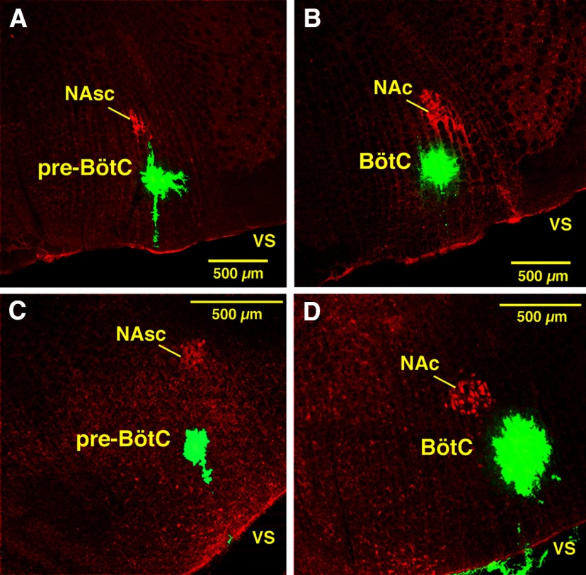
Figure 2.
Confocal microscopic images of histologic sections illustrating post hoc validation of microinjection sites marked by fluorescent microspheres (green) in the pre-BötC or BötC regions in fixed sections from anesthetized adult rat in vivo preparations and juvenile rat in situ brainstem–spinal cord preparations. A, B, Coronal sections (30 µm thick) of fixed tissue at the level of pre-BötC (A) and BötC (B) from in vivo preparations. C, D, Coronal sections (30 µm) of fixed tissue at pre-BötC (C) and BötC (D) levels from juvenile rat in situ brainstem–spinal cord preparations. Subdivisions of nucleus ambiguus (NAc, compact subdivision; NAsc, semi-compact subdivision), labeled with ChAT antibody (red), provide regional landmarks for pre-BötC (ventral to NAsc) and BötC (ventral to NAc) levels of the medulla. Each image is taken from serial coronal histologic sections obtained from experiments targeting these regions. Labeling with microinjected solution of fluorescent microbeads, with varying extent of local spread, indicates the approximate center of the drug microinjection sites in these examples. vs, ventral surface.

