
Figure 2.
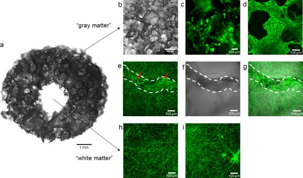
Neuronal compartmentalized outgrowth in the 3D brain-like tissue model. A) Macroscopic view of 3D bioengineered brain tissue, scale bar 1 mm; b) Macroscopic view of silk sponge showing the high porosity of the scaffold; c) Cell distribution upon attachment to the scaffold at day 1 of culture; d) Lack of collagen in the scaffold will result in no network formation due to the outgrowth of neurites on the surface of the silk (day 7); e) Neurons rapidly grow neurites into the surrounding collagen gel (day 7); f) Silk scaffold; g) Overlay image of neuronal projections and silk (e) and f), the white dotted lines indicate the silk scaffold on which cell bodies are present (red arrows). The cell bodies are distinctly visible as bright green foci; h) Representative image of neuronal network formed at day 7 in the central collagen window; i) Neuronal network with cell foci in sample with insufficient washing steps before collagen embedding. Cells visualized with anti-TUJ1 antibody fluorescently labeled with Alexa-488 followed by confocal microscopy. b-i scale bar 100 μm.