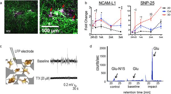
Figure 4.
Functional evaluation of the constructs. a) Immunostaining of 1 week old culture shows intimate localization of astrocytes (GFAP-magenta) in the direct proximity of neuronal cell bodies (TUJ-1-green). Scale bar 100 μm; b) Gene expression of neural cell adhesion molecule L1 (NCAM-L1) and synaptosomal-associated protein 25 (SNP-25) over 3 weeks of culture (red line) corresponding to neural network development over time. As a control the 3D brain like constructs are compared against regular 2D cultures (black line) and 3D dispersion cultures in collagen (blue line). Data are means ± SD. For the 2D and collagen groups, n = 3 replicates per time point per GOI. For the 3D scaffold group, n = 3, except for the 24-h and 1-wk time points, where n = 2, the 2-wk time point, where n = 2 for SNP- 25, and the 3-wk time point, where n = 2 for NCAM- L1 and growth-associated protein 43 and n = 1 for SNP- 25. Asymmetric error bars show maximum and minimum fold change. Two-way ANOVA with Bonferroni posttests: 2D vs. Col: *P <0.05; **P < 0.001; 2D vs. 3D: †P < 0.05; ‡P < 0.001; Col vs. 3D: §P < 0.05; c) Local field potential measurement. Spontaneous spiking activity is observed in our cultures, which can be diminished with 20 μM TTX; d) Injury-triggered glutamate (Glu) release. Glu peaks (arrow) at a retention time of ~21 min. Representative LC/MS detection traces of the internal control Glu-N15 and the Glu level at the baseline and after impact. The panels 4b-4d were previously published in Tang-Schomer, M. D. et al. Bioengineered functional brain-like cortical tissue. Proc. Natl. Acad. Sci. U.S.A. 111, 13811-13816 (2014).

