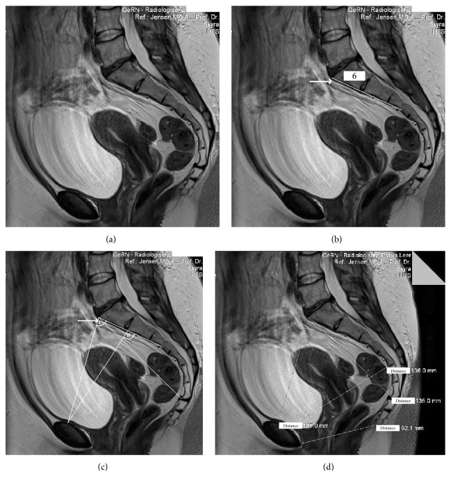
Figure 1.
MRI of the maternal pelvis (I.H.). (a) This MRI documents a “long pelvis” [2] with assimilated (fused) lumbar vertebral body to form a sacral bone with an excessive vertebral body (arrow). (b) The “long pelvis” has 6 vertebral bones instead of the normal 5. In addition, the 6 sacral bones in the “long pelvis” fail to be concave; rather they form a straight plane (indicated by the straight line), which reduces the capacity of the mid-pelvis. (c) The pelvic inlet angle is < 90° (angle I) in a “long pelvis” [2]. (d) The distance from the top of the 6th (excessive) vertebral body to the end of the sacral bone in this “long pelvis” [2] is prolonged ((d), distance 135 mm) (3-Tesla, T1w TSE triplanar sequence).

