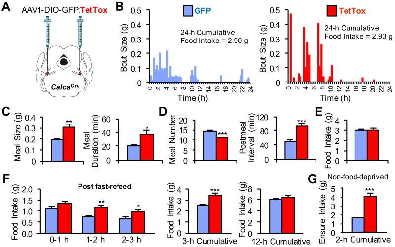
Figure 2. Functional Inactivation of CGRPPBel Neurons Disrupts Control of Meal Size.
(A) Bilateral delivery of AAV carrying Cre-dependent GFP:TetTox into the PBel of CalcaCre mice.
(B) Representative bout-size recordings from individual TetTox and GFP control mice during a 24-h recording period.
(C-E) Group-wide, meal-pattern analysis (n = 9 per group) was obtained from bout size recordings using set meal parameters. TetTox inactivation of CGRPPBel neurons increased the size and duration of meals (C) but was accompanied by a decrease in meal frequency (D) such that overall food intake was unaltered (E).
(F) Food intake measured following a 24-h food deprivation (n = 11 per group).
(G) Intake of a palatable liquid diet (Ensure) from non-food-deprived mice given 2 h ad libitum access during the light cycle (n = 11 per group). All data shown are means ± s.e.m. * P < 0.05; ** P < 0.01; *** P < 0.001. Statistical analysis was performed with an unpaired student’s t-test (C-D and G) and two-way ANOVA followed by Bonferroni’s post-hoc test (F). See also Figure S2.

