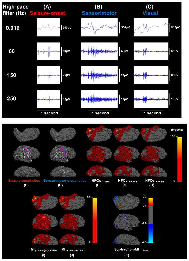
Figure 4. Spatial profiles of the rates of high-frequency oscillations (HFOs) and modulation index (MI) in patient #11.
HFO events at (A) seizure-onset, (B) sensorimotor, and (C) visual sites. (D) Seizure-onset sites: red circles. (E) Sensorimotor-visual sites: blue circles. (F) Rate of HFOs>80Hz (event/min). (G) Rate of HFOs>150Hz. (H) Rate of HFOs>250Hz. (I) MI(>150Hz)&(0.5–1Hz). (J) MI(>150Hz)&(3–4Hz). (K) Subtraction-MI>150Hz co-registered to MRI (SMICOM). Subtraction-MI>150Hz was defined as subtraction of MI(>150Hz)&(0.5–1Hz) from MI(>150Hz)&(3–4Hz). The highest value was noted in the seizure onset site, whereas nonepileptic sensorimotor-visual sites were associated with negative values.

