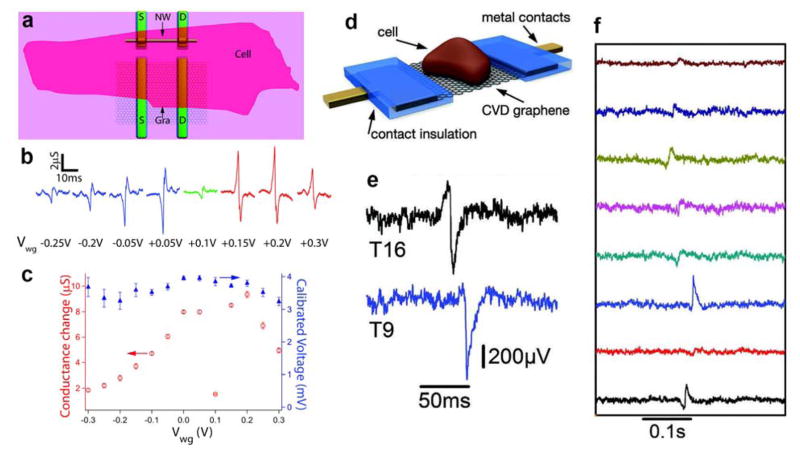
Figure 20.
Extracellular recording using graphene FETs. (a) Schematic illustrating cardiomyocyte cell interfaced to graphene- and SiNW-FET devices. (b) Recorded extracellular spikes versus gate potential. (c) Summary of the gate potential versus conductance change and calibrated voltage. Reprinted with permission from Ref. 544. Copyright 2010 American Chemical Society. (d) Schematic of a cell on a graphene-FET. (e) Typical two- and one-side peaks observed for different transistors. (f) Simultaneous current recordings from eight transistors in one FET array over hundreds of milliseconds. Reprinted with permission from Ref. 545. Copyright 2011 John Wiley & Sons, Inc.

