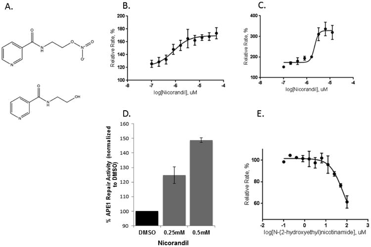
Figure 1.

Effect of nicorandil on APE1 activity. (A) Chemical structures for nicorandil (top) and the denitrated metabolite, N-(2-hydroxyethyl)nicotinamide (bottom) are shown. Increasing concentrations of nicorandil stimulate wild-type (B) and R177A APE1 (C) endonuclease activity. A nicorandil titration assay was carried out at 0.1 nM wild-type APE1 or 0.0125 nM R177A APE1 with 10 nM substrate in the presence of 1% DMSO. The concentration range of nicorandil was varied from 0.1 to 50 μM for wild-type APE1 and 0.1 to 12.5 μM for R177A APE1 with triplicate measurements for each concentration. The rates in relative fluorescent units were calculated by normalizing to the rate of 1% DMSO control in (B, C, D, and E). The curves were fit to a four-parameter dose response equation in Graph Pad. The EC50 is 0.82 μM for wild-type APE1 and 2.2 μM for R177A APE1. (D) Effect of nicorandil on APE1 activity from DRG cell extracts. Nicorandil was added directly to untreated DRG cell extract at the concentrations shown and assayed for APE1 endonuclease activity as previously described. Three independent experiments were performed with triplicate measurements for reactions including a total volume of 100 μL with 50 nM substrate and 3.3 μg extract in 2 % DMSO. Reaction mixtures included the same buffering components as in (B). (E) The denitrated metabolite of nicorandil, N-(2-hydroxyethyl)nicotinamide, inhibits the activity of APE1. Using the same kinetic fluorescent assay as described in Figure 1B, the effects of increasing the concentration of N-(2-hydroxyethyl)nicotinamide on APE1 activity is shown. An IC50 of 65 μM was determined by using a four parameter logistics curve in non-linear regression as implemented in GraphPad Prism.
