
Figure 4. Overexpression of miR-1343 reduces canonical TGF-β signalling.
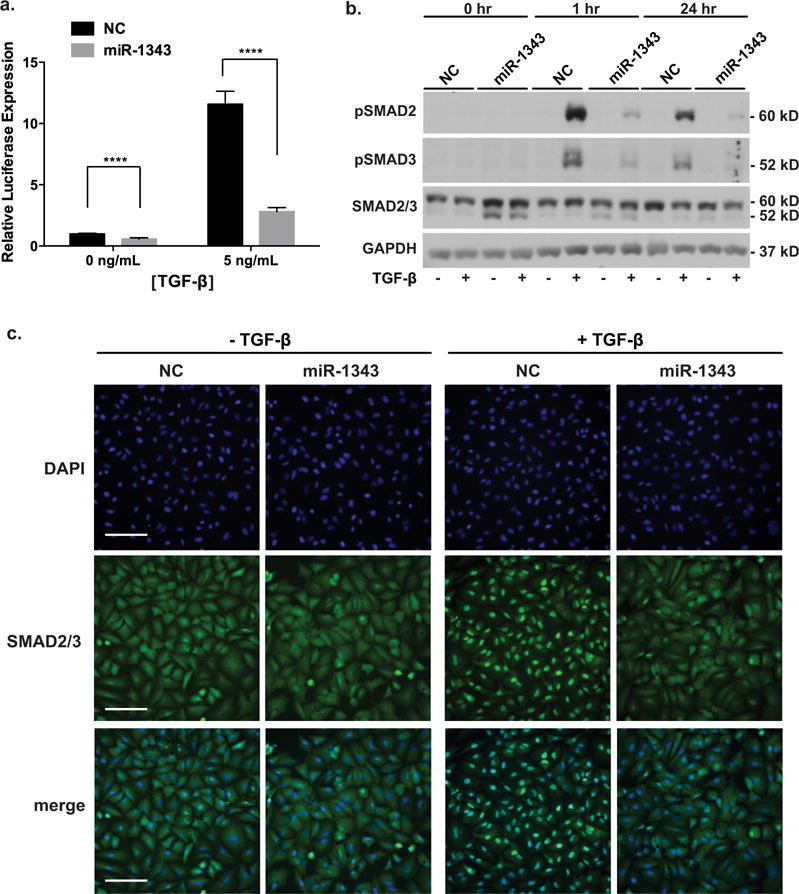
(a) Luciferase assay of A549 cells transiently transfected with the p3TP-lux vector and either miR-1343 or NC miRNA. Cells were treated with TGF-β1 (5 ng/ml) or vehicle control (0 ng/ml), 48 h after transfection, for 24 h. Luciferase values were normalized to pMIR-β-galactosidase levels and expressed relative to NC treated with vehicle. n = 3; ****P ≤ 0.0001. (b) miR-1343 represses phosphorylation of SMAD2/3. Western blot of lysates from A549 cells transiently transfected with miR-1343 or NC miRNA and treated with TGF-β1 (5 ng/ml, +) or vehicle control (−) for the indicated period of time. Blots were probed with antibodies specific for pSMAD2 [phosphorylated (active) SMAD2], pSMAD3 [phosphorylated (active) SMAD3] and total SMAD2/3; slower migrating species is SMAD2 and faster is SMAD3. GAPDH was the loading control. (c) miR-1343 inhibits nuclear localization of pSMAD2/3. Representative images of immunofluorescence in A549 cells transiently transfected with miR migrating species-1343 or NC miRNA and treated with TGF-β1 (50 ng/ml, +) or vehicle control (−) for 1 h. Green fluorescence shows total SMAD2/3 and blue is DAPI nuclear counterstain. Merge illustrates total SMAD2/3 plus DAPI. Scale bar = 100 μm.