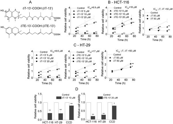
Figure 1. The structures of δT-13’-COOH and δTE-13’-COOH (A); The effect of 13’-COOHs and tocopherols on cell viability of human colon HCT-116 (B) and HT-29 (C) cancer cells; The relative anti-proliferation effect (24-h incubation) of 13’-COOHs on cancer cells vs. human normal CCD841CoN (CCD) colon epithelial cells (D).
Relative cell viability was measured by MTT assay after cells were treated with 13’-COOHs or tocopherols at indicated concentrations and times compared with DMSO controls. IC50s are the concentrations that caused 50% decrease in cell viability after 24-h incubation. The data are mean SD from at least three independent experiments. *p < 0.05 and **p < 0.01 indicate a significant difference between treated and DMSO-control cells. δT-13’-COOH and δTE-13’-COOH are abbreviated as δT-13’ and δTE-13’, respectively.

