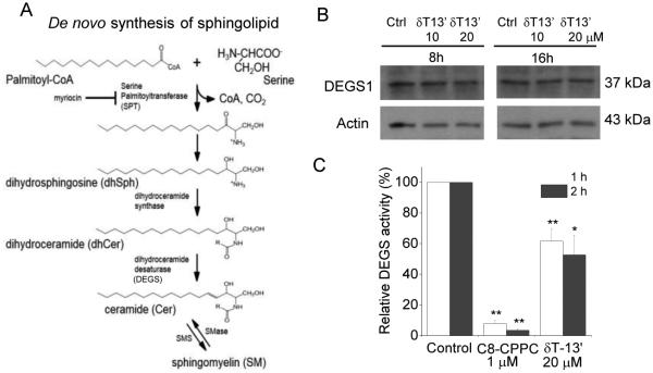
Figure 4. δT-13’-COOH (δT-13’) inhibited DEGS activity but not protein expression.
A: The de novo biosynthesis pathway of sphingolipids (SMS: sphingomyelin synthase; SMase: sphingomyelinase). B: HCT-116 cells were treated with δT-13’-COOH at 10 or 20 μM for 8 or 16 h and the effect on DEGS-1 expression was detected by Western blotting. C: HCT-116 cells were treated with DMSO (control), δT-13’-COOH at 20 μM or C8-CPPC at 1 μM for 1 or 2 h. Cells were collected and homogenized. The homogenates were added with NADH and C8:0-dhCer as a substrate for DEGS. After 1 h incubation at 37 C, the production of C8:0-sphingolipids were analyzed by LC-MS/MS. The data are mean SEM of three independent experiments. *p < 0.05 and **p < 0.01 indicate significant differences between DMSO-control cells and 13’-COOH or CPPC-treated cells.

