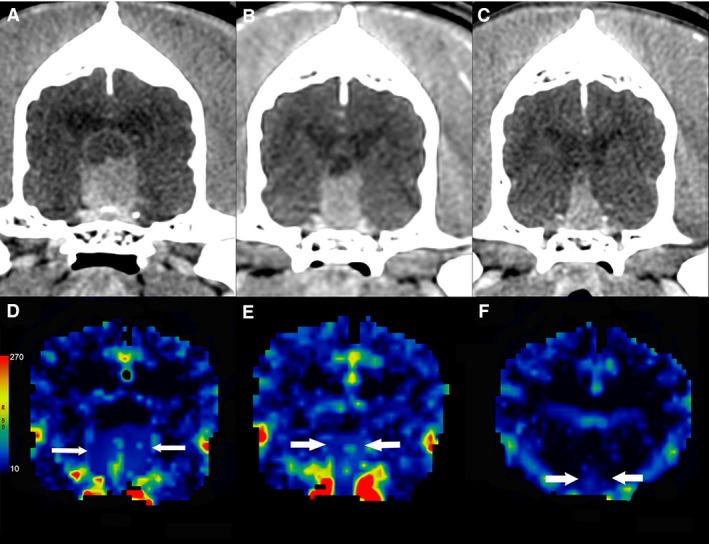
Figure 2.

Contrast‐enhanced transverse CT images of a dog with a pituitary tumor treated with stereotactic radiosurgery. The mass decreases in size from the baseline images (A) through the first (B) and second (C) rechecks, from left to right with corresponding blood volume maps (mL/100 g) (D, E, F).
