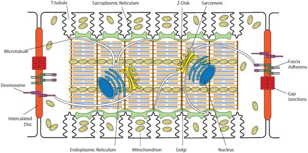
Figure 1. Organization of an adult ventricular cardiomyocyte.
Intercalated discs are located at the longitudinal sides of each ventricular cardiomyocyte to mediate cell-to-cell mechanical and electrical coupling. Mechanical coupling of cardiomyocytes is achieved through desmosomes and fascia adherens complexes. Gap junctions are primarily responsible for electrical coupling of the cardiomyocytes. T-tubules, which are rich in voltage-gated L-type calcium channels, are positioned closely to the sarcoplasmic reticulum, the internal calcium store. Sarcomeres form myofibrils, which are responsible for cardiomyocyte contraction upon intracellular calcium release. The Golgi apparatus and microtubules serve as the “loading dock” and “highways”, respectively, to deliver ion channels to specific subdomains on the plasma membrane. Mitochondria provide the energy needed for the contraction of cardiomyocytes.

