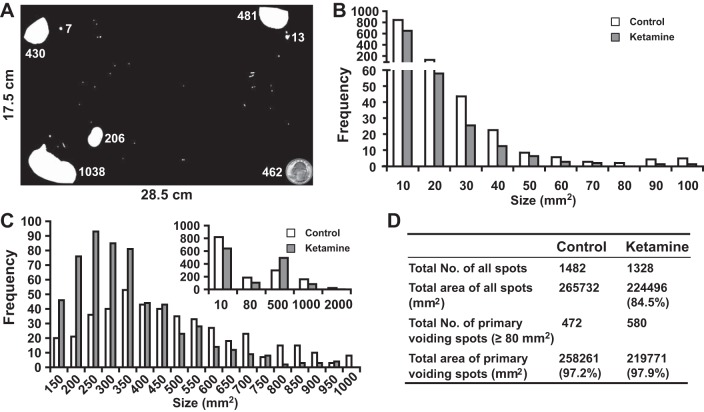
Fig. 2.
Voiding spot assay. All voiding spots from control (n = 10) and ketamine (n = 10)-treated groups taken between weeks 2 and 12 were imaged and measured for their spot size/area (in mm2). Spot areas ranged from 1.5 to 2,250 mm2. Spots having an area of <1.5 mm2 were discarded. A: representative filter paper with dimensions of 28.5 × 17.5 cm. The white areas on the filter paper are voiding spots, and the adjacent number indicates the spot area (in mm2). The quarter dollar coin at the bottom right corner has a surface area of 462 mm2, which provides a comparison with the actual size of the voiding spots. All spots were then sorted according to their size. B: frequency distribution chart of all voiding spots with a size of <100 mm2. The x-axis indicates the size of the voiding spots, and the number below the axis indicates the spot size range. For example, 10 indicates a spot size range from 1.5 (low threshold) to 10 mm2 and 20 indicates a spot size range from 10 to 20 mm2 in this chart. We noticed that many spots were <10 mm2, and the frequency of spot size decayed gradually with the increase of size until it reached 0 or near 0 at a spot size of 70–80 mm2. At a spot size of larger than 80 mm2, the frequency increased. C: frequency distribution chart of all voiding spots with a size ranging from 100 to 1,000 mm2. The x-axis indicates the size of the voiding spots, and the number below the axis indicates the spot size range. For example, 150 indicates a spot size range from 101 to 150 mm2 and 200 indicates a spot size range from 151 to 200 mm2. Please note that the x-axis of the inset chart has a different scale, for example, 500 indicates a spot size ranging from 81 to 500 mm2. Voiding spots from the ketamine-treated group were more clustered around the size of 201–350 mm2, whereas the range of voiding spots from the control group was broader. The inset in C shows an overall frequency distribution chart of all voiding spots (from 1.5 to 2,250 mm2). According to these frequency distribution charts, we attributed voiding spot sizes of >80 mm2 as primary voiding spot (PVSs), as shown in D, which accounted for ∼98% of the total urine area. It was also clear that ketamine-treated group had more PVS numbers, indicating voiding frequency.

