Fig. 7.
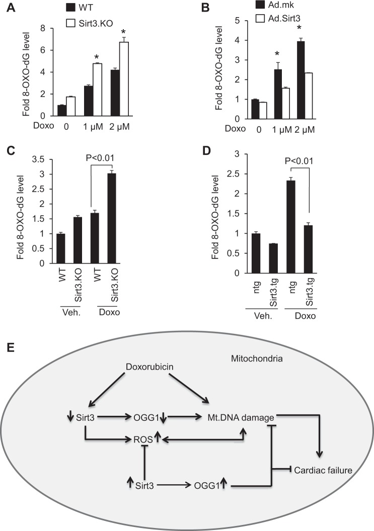
Sirt3 overexpression decreases 8-oxo-dG levels. A: primary cultures of mouse cardiac fibroblasts obtained from WT and Sirt3.KO mice were treated with Doxo for 24 h and 8-oxo-dG content in total DNA was measured. *Significantly higher (P < 0.01) compared with cells from WT mice. B: primary cultures of cardiomyocytes were infected with adenovirus expressing SIRT3 or mock adenovirus (Ad.mk). Following 24 h of infection, cells were treated with different doses of Doxo as indicated and 8-oxo-dG content in total DNA was measured. *Significantly higher (P < 0.01) compared with cells overexpressed with Sirt3. C: 8-oxo-dG content in the DNA of whole heart of WT and Sirt3.KO mice treated with saline or Doxo. D: 8-oxo-dG content in the DNA of whole heart of ntg control and Sirt3.tg mice treated with saline or Doxo. All values are means ± SE; n = 5. E: model illustrating how Sirt3 protects the heart from Doxo-induced cardiac injury. Doxo treatment downregulates Sirt3 levels in the heart resulting in decreased levels of OGG1 and increased levels of ROS in mitochondria, both contributing to mtDNA damage. Doxo can also cause DNA damage by directly binding to DNA. Activation of Sirt3 leads to increased OGG1 levels and decreased ROS production, thereby mitigating mtDNA damage by decreasing the oxidative damage as well by increasing the efficiency of DNA repair. These changes together with the ability of Sirt3 to promote mitochondrial fusion enhance the overall health of mitochondrial population, and thereby protecting cardiac myocytes from death and the development of cardiac failure.

