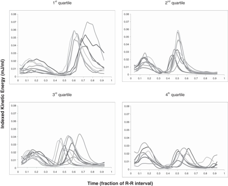
Fig. 2.
KE in the left ventricle (LV) according to age. These images show KE indexed to the ventricular volume at each time point during the cardiac cycle. Indexing KE to the volume of blood at that time allowed comparison between subjects of different sizes. The cardiac cycle was divided into fractions of the R-R interval. Top left: 1st age quartile; top right: 2nd age quartile; bottom left: 3rd age quartile; bottom right: 4th age quartile.

