Fig. 1.
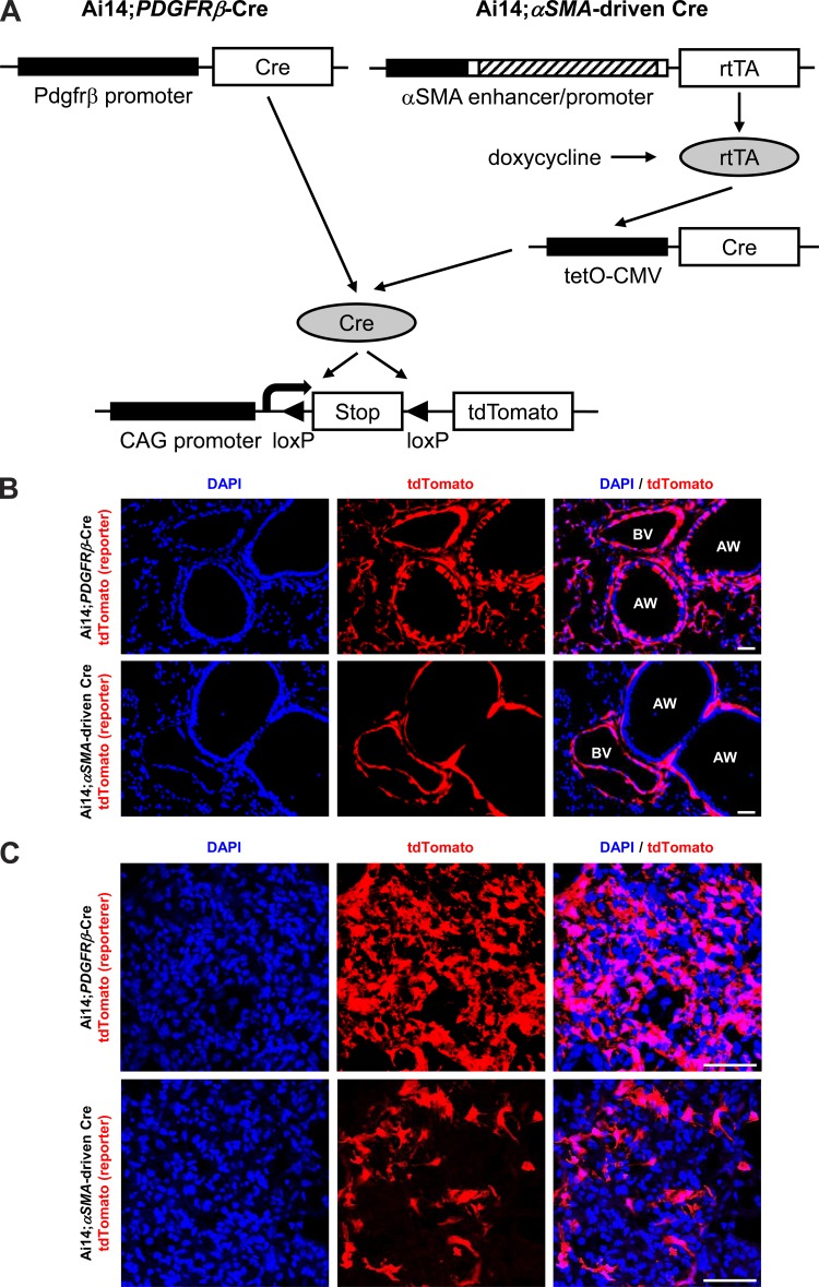
Comparison of tdTomato expression pattern between Ai14;PDGFRβ-Cre and Ai14;αSMA-rtTA;tetO-Cre mice in normal and fibrotic lungs. A: schematic of reporter mice generated by crossing Ai14 mice (29) with either PDGFRβ-Cre (12) or αSMA-driven Cre mice (9, 37). The αSMA enhancer/promoter construct contained that consisted of ∼1 kb of the 5′-flanking region (black box), the transcription start site, the 48-bp exon 1 (white box), the ∼2.5-kb intron 1 (hatched box), and the 15-bp exon 2 (white box) from the mouse αSMA gene (49). B: immunofluorescence micrographs of 14-day water-treated lung sections from the indicated reporter mice. AW, airway; BV, blood vessel. C: immunofluorescence micrographs of 14-day bleomycin-treated lung sections from the indicated reporter mice. Panels at left show DAPI (blue), panels in middle show endogenous tdTomato (red), and panels at right show merged images. Scale bar, 100 μm.

