Fig. 3.
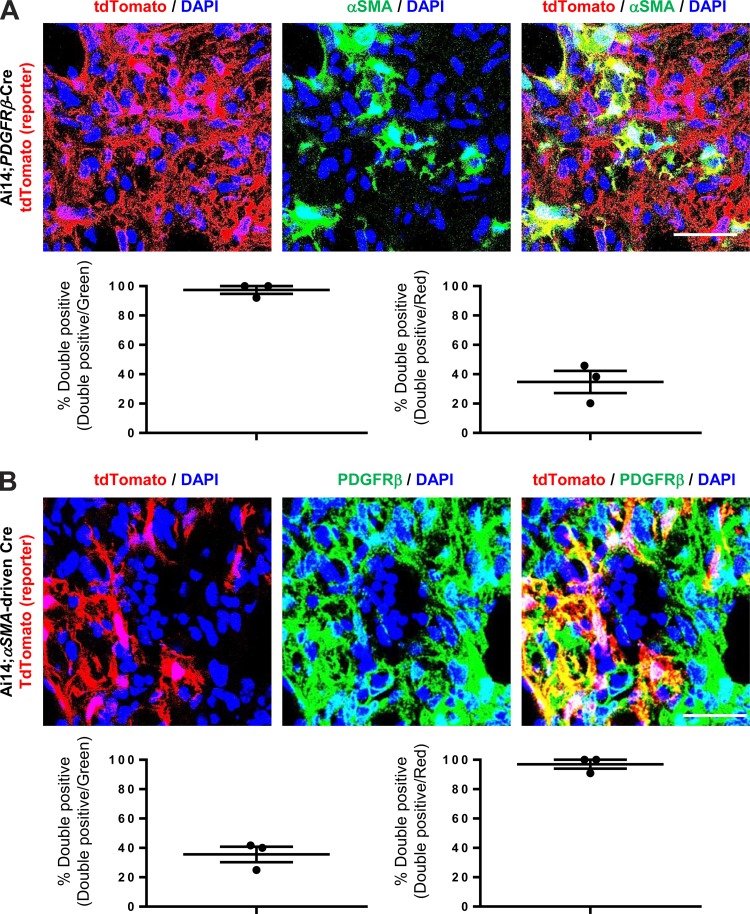
αSMA-expressing cells are only a subpopulation of PDGFRβ-expressing cells within the fibrotic lung interstitium. A: immunofluorescence micrographs and image quantification of lung sections from 14-day bleomycin-treated Ai14;PDGFRβ-Cre mice, which were costained with αSMA antibody. Panel at left shows DAPI (blue) and endogenous tdTomato (i.e., PDGFRβ) (red), panel in middle shows DAPI (blue) and αSMA staining (green), and panel at right shows the merged image. B: immunofluorescence micrographs and image quantification of lung sections from 14-day bleomycin-treated Ai14;αSMA-rtTA;tetO-Cre mice, which were costained with PDGFRβ antibody. Panel at left shows DAPI (blue) and endogenous tdTomato (i.e., αSMA) (red), panel in middle shows DAPI (blue) and PDGFRβ staining (green), and panel at right shows the merged image. Scale bar, 50 μm.

