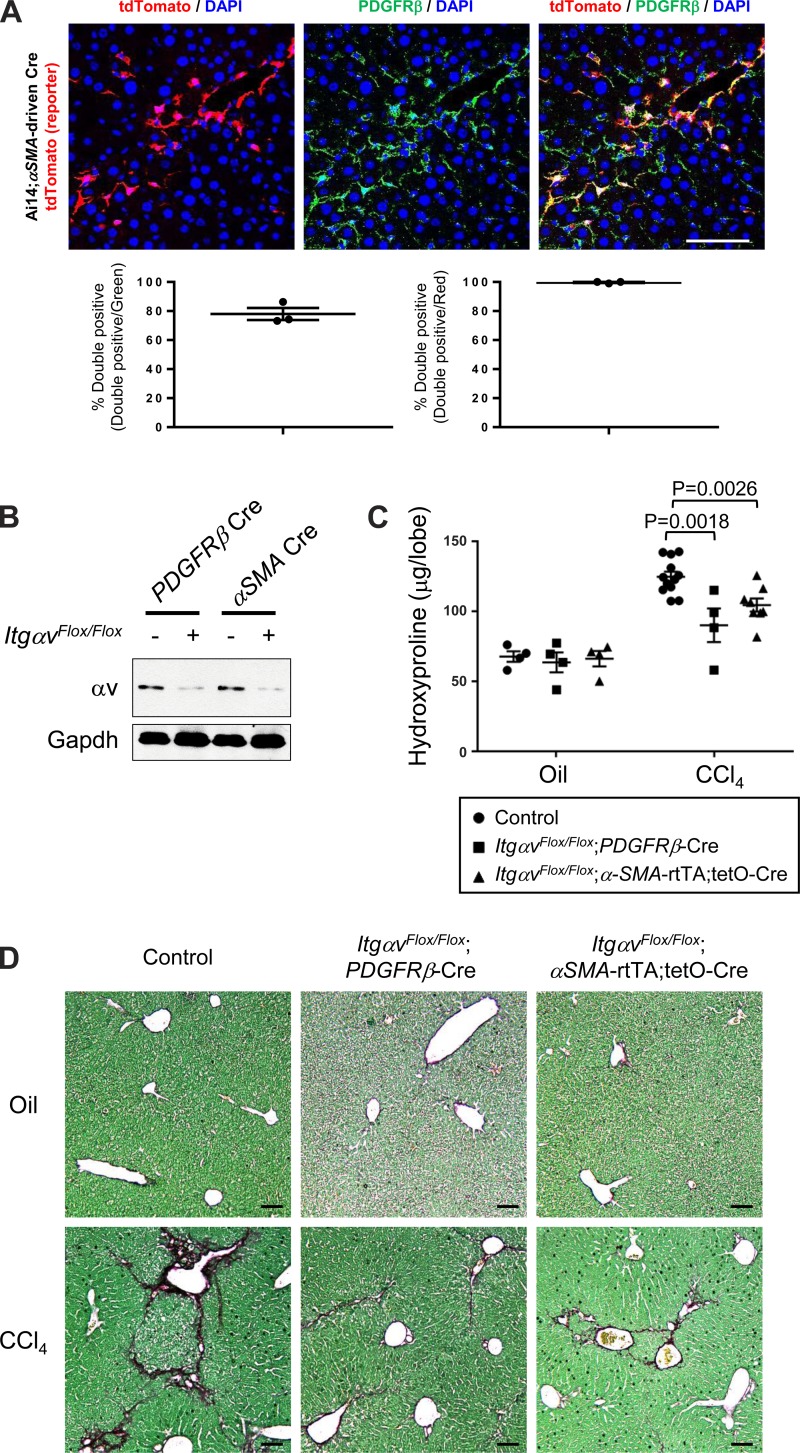
Fig. 5.
αSMA-directed deletion of αv integrins protects against CCl4-induced hepatic fibrosis. A: immunofluorescence micrographs and image quantification of liver sections from 3-wk CCl4-treated Ai14;αSMA-rtTA;tetO-Cre mice, costained with PDGFRβ antibody. Panel at left shows DAPI (blue) and endogenous tdTomato (i.e., αSMA) (red), panel in middle shows DAPI (blue) and PDGFRβ staining (green), and panel at right shows the merged image. Scale bar, 100 μm. B: immunoblotting of expression of αv integrins in cells targeted by PDGFRβ-Cre or αSMA-driven Cre. Mice were treated with CCl4 for 3 wk, and tdTomato-positive cells were purified by FACS, followed by immunoblotted with anti–αv integrin antibody. C: hydroxyproline assay of liver lysates. D: picrosirius red staining of liver from mice treated with olive oil (control vehicle) or CCl4 twice a week for 6 wk. Data are means ± SE. P value, Student's t-test. Scale bar, 200 μm.

