Fig. 3.
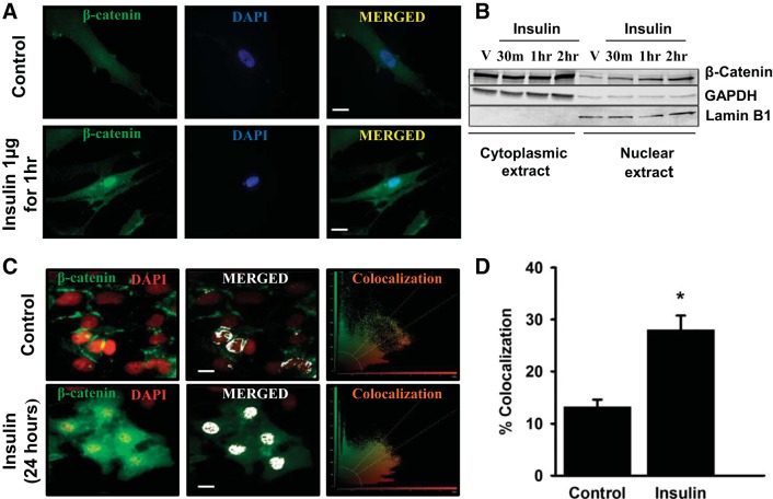
Insulin induces β-catenin nuclear localization in ASM and BEAS2B cells. The effect of insulin on subcellular localization of β-catenin was investigated in both transformed human bronchial epithelial (BEAS2B) and primary human airway smooth muscle (ASM) cells. Insulin (1 μg)-induced β-catenin translocation to nucleus, in ASM cells, was confirmed by fluorescence imaging after 1 h (A) and immunoblotting of β-catenin in cytoplasmic and nuclear extracts before insulin treatment (V) and at different time points [30 min (30 m), 1 h, and 2 h] posttreatment (B). β-Catenin translocation to nucleus in BEAS2B cells was confirmed by measuring colocalization rates of fluorescent markers represented as green (β-catenin) and red (nucleus). Left, merged fluorescent image; middle, colocalized regions (white); right, scatterplot of the acquired image processed by the LAS AF software (Leica). The colocalization rate is the percentage of colocalization extent calculated from the ratio of the areas of colocalizing fluorescent markers and that of image foreground (C). Bar plot showing the mean colocalization rates corresponding to control and insulin-treated BEAS2B cells (D). Magnification ×63; scale 20 μm. Data represent means ± SE, n = 6, *P ≤ 0.05, compared with respective controls. GAPDH, glyceraldehyde-3-phosphate dehydrogenase; V, vehicle.

