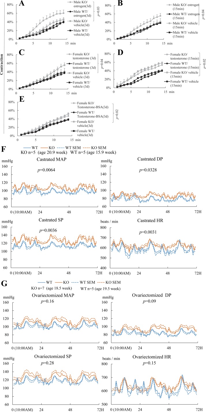
Fig. 5.
Estrogen enhances but testosterone suppresses and VSMC contractility and hence BP in the absence of EFNB3. VSMCs were cultured in medium containing charcoal-stripped FCS for 3–4 days. Means ± SE of percentage contraction of more than 15 cells per group are registered. Statistically significant differences were assessed by ANOVA followed by post hoc examination. P values between groups with significant differences are indicated. All experiments were conducted 3 times independently. Data from representative experiments are presented. A: long-term estrogen treatment augments contractility of VSMCs from KO but not WT mice. VSMCs from WT and KO males were cultured for 3–4 days in the presence of 17β-estradiol (100 pg/ml) or vehicle and then stimulated with PE (20 μM). Cell contractility was registered. B: short-term estrogen treatment augments contractility of VSMCs from KO but not WT male mice. VSMCs from male KO or WT mice were cultured for 3–4 days and then stimulated with 17β-estradiol (100 pg/ml) or vehicle for 15 min. In the same last 15 min of culture, PE (20 μM) was also added and VSMC contractility was recorded. C: long-term testosterone treatment suppresses contractility of VSMCs from female KO but not WT mice. VSMC from WT and KO females were cultured for 3–4 days in the presence of 5α-dihydrotestosterone (6.49 ng/ml) or vehicle and then stimulated with PE (20 μM). VSMC contractility was recorded. D: short-term testosterone treatment has no effect on contractility of VSMCs from female KO or WT mice. VSMCs from female KO or WT mice were cultured for 3–4 days and then stimulated with 5α-dihydrotestosterone (6.49 ng/ml) or vehicle for 15 min. In the last 15 min of culture, PE (20 μM) was also added and VSMC contractility was recorded. E: cell membrane-impermeable testosterone-3-(O-carboxymethyl)-oxime-BSA has not effect on contractility of VSMCs from female KO or WT male mice. VSMCs from female KO or WT mice were cultured for 3–4 days in the presence of cell membrane-impermeable testosterone-3-(O-carboxymethyl)-oxime-BSA (1.1 μg/ml) and then stimulated with PE (20 μM). VSMC contractility was recorded. F and G: castrated male KO mice are hypertensive and ovariectomized female KO mice are normotensive. BP and HR of castrated male (F) and ovariectomized female (G) KO and WT mice were measured by radiotelemetry as described in Fig. 2. Telemetry transmitters were implanted in the mice, and after at least 1 mo, they were castrated or ovariectomized. Telemetry was conducted 3 wk after gonadectomy. Number per group and the mean ages of the mice at the time of BP measurement are indicated. The BP and HR of all mice were measured for 72 h. The 72-h values were used for statistical analysis (ANOVA). P values are indicated.

