Fig. 2.
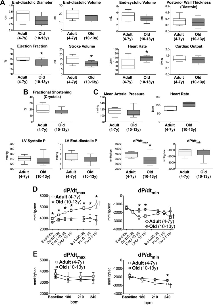
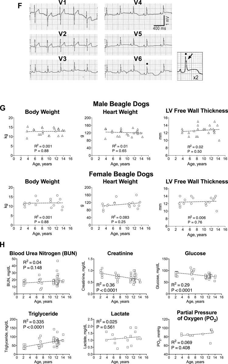
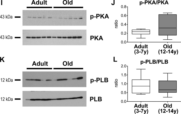
Aging negatively interferes with cardiac function. A: echocardiographic parameters in aging dogs; data are shown as median and interquartile ranges (adult: n = 6, 5.5 ± 0.6 yr; old: n = 8, 11.9 ± 0.4 yr). *P < 0.05 vs. adult; y, yr of age. B: radial left ventricular (LV) contractility in aging dogs; data are shown as median and interquartile ranges (adult: n = 5, 5.9 ± 0.6 yr; old: n = 8, 11.8 ± 0.4 yr). *P < 0.05 vs. adult. C: hemodynamic parameters in aging dogs; data are shown as median and interquartile ranges (adult: n = 6, 5.5 ± 0.6 yr; old: n = 9, 12.0 ± 0.4 yr). *P < 0.05 vs. adult. D: maximal rate of LV contraction and relaxation in aging dogs following infusion of dobutamine (Dobt, dosage referred to kg of body wt per minute of infusion) and bolus of isoproterenol (Iso, dosage referred to kg of body wt) are shown as mean ± SE (adult: n = 6, 5.5 ± 0.6 yr; old: n = 6, 12.5 ± 0.4 yr). *P < 0.05 vs. adult; †P < 0.001 within the same group E: effects of overdrive pacing are shown as mean ± SE (adult: n = 6, 5.5 ± 0.6 yr; old: n = 5 12.0 ± 0.6 yr). *P < 0.05 vs. adult; †P < 0.01 within the same group. F: ECGs from precordial leads obtained in a dog at 11 yr of age. Notched R waves are present in V6. Arrow points to the notched R wave in the magnified trace. G: gross anatomical parameters in aging dogs. Body weight before death and heart weight and LV free wall thickness measured in the explanted organ (male: n = 21; female: n = 18). Data are fitted with linear regression. R2 and P value for each fitting are reported. H: biochemical parameters of blood samples obtained from the jugular vein, with the exception of lactate and partial pressure of oxygen, measured in blood collected from the coronary sinus. Data are fitted with linear regression. R2 and P values for each fitting are reported. I–L: Western blots analysis for total and phosphorylated PKA and phospholamban (PLB) in the LV myocardium of aging dogs. Quantitative data for p-PKA/PKA ratio (J) in the LV myocardium of adult (n = 11, 5.2 ± 0.4 yr) and old (n = 12, 12.9 ± 0.2 yr) beagle dogs are shown as median and interquartile ranges. Quantitative data for p-PLB/PLB ratio (L) in the LV myocardium of adult (n = 10, 5.2 ± 0.5 yr) and old (n = 12, 12.9 ± 0.2 yr) beagle dogs are shown as median and interquartile ranges.


