Fig. 4.
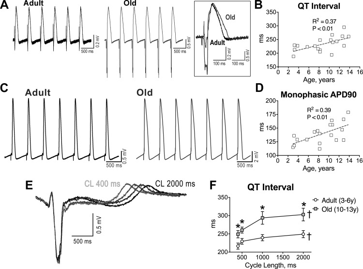
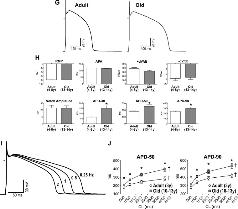
Aging results in prolongation of the electrical recovery of myocardium and myocytes. A: pseudo-ECGs in the perfused adult and old LV myocardium stimulated at 2 Hz. Superimposed traces are shown in the inset. B: quantitative measurements of QT interval duration in the myocardium of beagle dogs (n = 19 muscles, 19 dogs) are fitted with linear regression. R2 and P values are reported. C: monophasic action potentials (MAPs) measured in adult and old LV tissue. D: quantitative measurements of duration of the action potential (AP) at 90% repolarization (APD90; n as in B) are fitted with linear regression. R2 and P values are reported. E: superimposed pseudo-ECGs obtained at different pacing cycle lengths (CL). F: data are shown as mean ± SE (adult: n = 11 muscles, 6 dogs, 3.8 ± 0.4 yr; old: n = 8 muscles, 7 dogs, 11.5 ± 0.4 yr). *P < 0.05 vs. adult; †P < 0.001 for various frequencies within the same group. G: APs of LV myocytes from adult and old dogs. H: data are shown as mean ± SE (adult: n = 22 cells, 8 dogs, 5.6 ± 0.4 yr; old: n = 10 cells, 5 dogs, 13.1 ± 0.1 yr). *P < 0.05 vs. adult. RMP, resting membrane potential; APA, AP amplitude. I: superimposed APs at various pacing frequencies in a LV myocyte from an adult dog. J: data are shown as mean ± SE (adult: n = 16 cells, 2 dogs, 3.2 ± 0 yr; old: n = 26 cells, 6 dogs, 10.9 ± 0.1 yr). *P < 0.05 vs. adult; †P < 0.001 for various frequencies within the same group.


