Fig. 5.
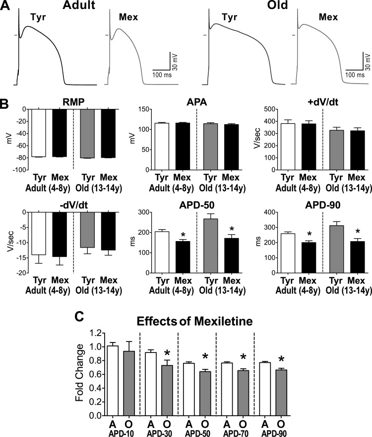
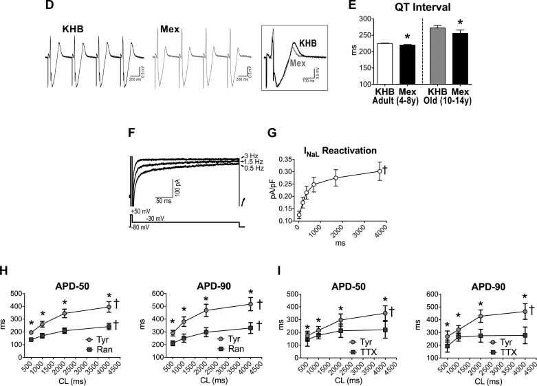
A: APs recorded in 1 adult and 1 old myocyte in Tyrode (Tyr) and after exposure to mexiletine (Mex; 10 μM). B: data are shown as mean ± SE (adult: n = 19 cells, 8 dogs, 5.2 ± 0.4 yr; old: n = 10 cells, 4 dogs, 13.1 ± 0.1 yr). *P < 0.001 vs. Tyr. C: comparison of the effects of late Na+ current (INaL) inhibition on AP duration between adult (A) and old (O) myocytes reported in B. data are shown as mean ± SE. *P < 0.01 vs. A. D: pseudo-ECG in the perfused LV myocardium of an adult dog. Traces in Krebs-Henseleit buffer (KHB) solution and after exposure to Mex (10 μM) are reported and superimposed in inset. E: data are shown as mean ± SE (adult: n = 6 muscles, 6 dogs, 6.5 ± 0.9 yr; old: n = 6 muscles, 4 dogs, 12.7 ± 0.5 yr). *P < 0.05 vs. KHB. F: superimposed traces of INaL (top traces) measured in voltage-clamp using a voltage-command protocol (bottom trace) at 0.5, 1.5, and 3 Hz, in a juvenile hound type dog myocyte. INaL was progressively reduced at higher frequencies. G: quantitative data for INaL reactivation in LV myocytes form juvenile (0.6–0.8 year old) hound type dogs are reported as mean ± SE (n = 29 cells, 4 dogs). †P < 0.001 for various frequencies. Amplitude of INaL is reported as positive value. H and I: data relative to effects of INaL inhibitors ranolazine (Ran; 10 μM) (H) and low doses of tetrodotoxin (TTX; 1 μM) (I) on AP profile of cardiomyocytes from old beagle dogs (11–13 yr of age) are shown as mean ± SE (ranolazine: n = 10 cells, 2 dogs 11 yr old; TTX: n = 5 cells, 2 dogs, 11–13 yr). *P < 0.05 vs. Tyr; †P < 0.001 for various frequencies within the same group.


