Fig. 6.
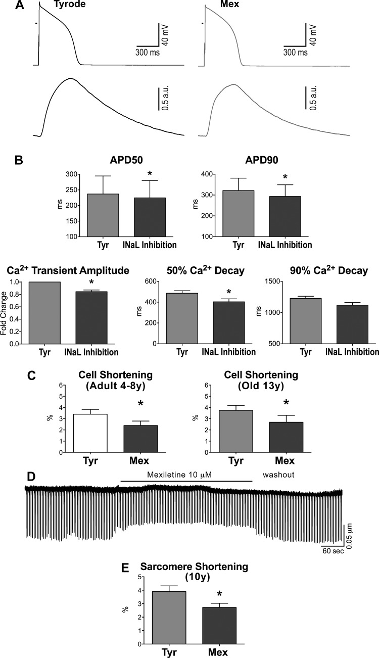
INaL has inotropic effects on cardiomyocytes. A: AP (top traces) and Ca2+ transients (bottom traces) recorded simultaneously in a myocyte from an old dog in Tyr and after exposure to the INaL inhibitor Mex (10 μM). B: quantitative data for AP and Ca2+ transient properties in old myocytes, in Tyr, and in the presence of INaL inhibition (10 μM Mex, n = 1, or 1 μM TTX, n = 3); data are shown as mean ± SE (n = 4 cells, 2 dogs, 11–14 yr old). *P < 0.05 vs. Tyr. C: data on contractile (cell shortening) properties in adult (left: n = 14 cells, 6 dogs, 6.3 ± 0.9 yr) and old (right: n = 6 cells, 2 dogs, 13.1 ± 0.3 yr) myocytes stimulated at 0.25–0.5 Hz in Tyr and following exposure to Mex (10 μM) are shown as mean ± SE. *P < 0.05. D: trace of sarcomere shortening for an old myocyte during the exposure to Mex and during the washout phase. E: data of contractility (sarcomere shortening) for old myocytes are shown as mean ± SE (n = 7 cells, 1 dog, 10 yr old). *P < 0.001 vs. Tyr.

