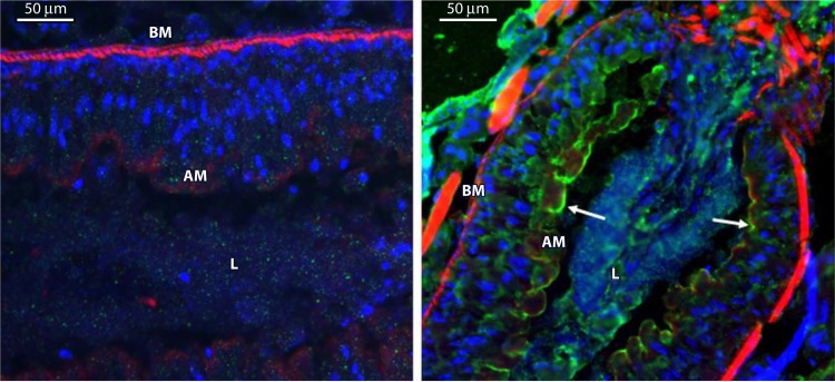
FIG 10.
Immunolocalization of Vip3Aa in midgut tissue sections after ingestion by S. frugiperda larvae. (Left) Control larvae. (Right) Larvae that ingested Vip3Aa. Nuclei were stained blue, and the apical and basal membranes were stained red. Binding of Vip3Aa to the apical membrane is shown in green. BM, basal membrane; AM, apical membrane; L, gut lumen. (Reprinted from reference 89.)

