Fig. 1.
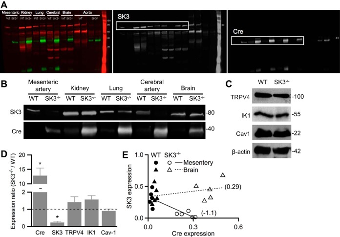
Lack of endothelial small-conductance Ca2+-activated potassium 3 (SK3) channels in tissue-specific SK3−/− mice. Western blot analysis performed on tissues obtained from homozygous floxed SK3 expressing endothelium-specific Cre-recombinase (SK3−/−) or wild-type (WT) mice. A: representative blots prepared from whole brain, lung, or kidney lysate, or lysates of aortas or cerebral or mesenteric arteries, were probed with anti-SK3 or anti-Cre recombinase antibodies. Color channels were separated to show SK3 (red) and Cre (green) expression in black and white. B: black and white representation of the indicated sections in A with enhancement of brightness/contrast. C: representative blots prepared from WT and SK3−/− mesenteric arteries were probed for transient receptor potential vanilloid 4 (TRPV4), IK1, and caveolin-1 (cav-1), and the expression densitometry analyses were normalized to β-actin signal. D: protein expression ratios in SK3−/− vs. WT. Asterisks (*) denote statistical significance using Wilcoxon rank test (P < 0.05) comparing SK3−/− and WT protein densitometry normalized to β-actin. Data are expressed as means ± SE; n = 5 mice from each genotype. E: expression of SK3 and Cre normalized to β-actin in WT (solid) and SK3−/− (empty) mesenteric artery (circle) or brain lysate (triangle). Linear regression fits (lines) and slopes (numbers in brackets) show negative correlation of SK3 and Cre in mesenteric arteries. Each point represents an animal; n = 5 animals from each genotype.

