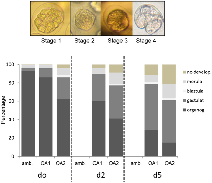
Figure 3. Upper - Inverted microscope images of the different egg-developmental stages.
Stage 1 (morula, cleavage), Stage 2 (blastula, cleavage), Stage 3 (late gastrulation), Stage 4 (organogenesis); Lower - Embryonic development after 72 h, expressed as the percentage of the different stages at ambient conditions (amb., pH 8.), low pH (OA1, pH 7.8), and extremely low pH (OA2, pH 7.6). The eggs were incubated from females exposed to 0, 2 and 5 days of OA treatment (d0, d2 and d5 respectively).

