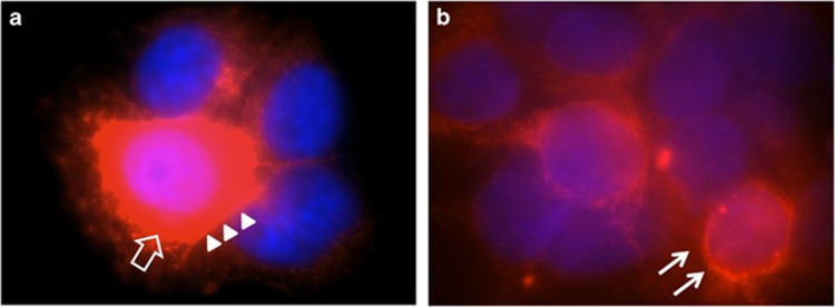
Figure 2.
Immunofluorescence staining using a primary antibody against RET upon transfections in HEK293 cells with: (a) pCMV-RET-WT and (b) pCMV-RET-p.(Pro270Leu). RET-WT staining shows strong signals in the cytoplasmic (empty arrow) and cell membrane (arrow heads), whereas the expression of RET-p.(Pro70Leu) is less pronounced and localized closer to the nucleolus/endoplasmic reticulum (arrow).

