Figure 5.
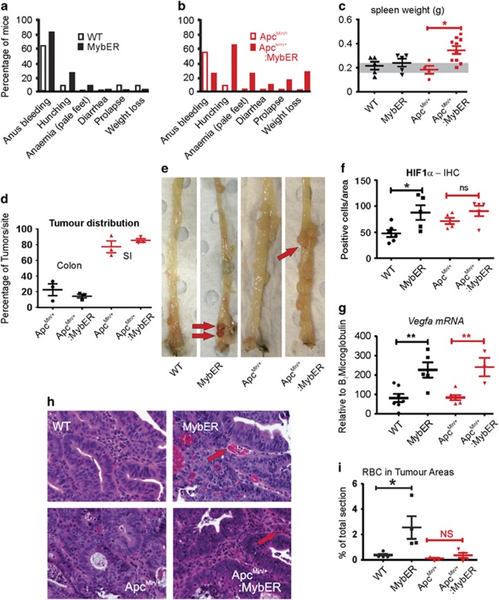
MybER-activation promotes CRC-associated clinical symptoms. (a) AOM-treated mice on a WT or (b) Apcmin/+ background were euthanized owing to a spectrum of symptoms. (c) Splenomegaly was significantly higher in Apcmin/+:MybER mice (grey zone indicates normal spleen weights in age-matched WT mice). (d) The distribution of tumors between small intestines and colon was similar in AOM treated Apcmin/+:MybER and Apcmin/+ mice. (e) Tumors arising on a MybER background were consistently redder indicative of an increase in red blood cells (RBC) (red arrows show red tumors). (f) The tumors were also significantly more hypoxic as shown by HIF1α IHC and (g) Vegfa mRNA expression was significantly higher in mice on a MybER background. (h–i) Although vascularization was not found to increase in MybER mice (see Supplementary Figure 8) the amount of RBCs in tumors on a WT background was most apparently increased (red arrows). Data presented for individual samples (n>5) with means ±s.e.m.; *P<0.05, two-sided t-test.

