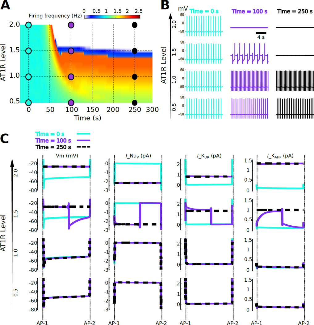
Fig. 1. Bifurcation of firing rate response to AngII as a function of AT1R level.
(A) Firing rate response to continuous 100 nM AngII application starting at time = 0 as a function of time for a population of cells exhibiting variation in AT1R level. The AT1R level was varied from 0.5 to 2 times the reference level. A narrow region of AT1R levels (~ 1.5×, blue) separates firing rate increases (<1.5×) from firing rate cessation (>1.5×). This narrow region of AT1R levels is referred to as a separatrix. (B) Membrane potentials are shown at three time points (t = 0, 100, and 250 sec) for four AT1R levels (0.5×, 1×, 1.5×, and 2×). (C) Inter-AP potentials and currents corresponding to the times and AT1R levels from panel B. Vm denotes membrane potential and Ix denotes the ionic current due to channel x. Only currents that showed detectable changes following AngII application are shown: INa, IKDR, and IKAHP. The data indicate that the separatrix occurs because increased AT1R signaling results in depolarization resulting from re-balancing of Na+ and K+ currents, with consequent abrogation of AP firing.

