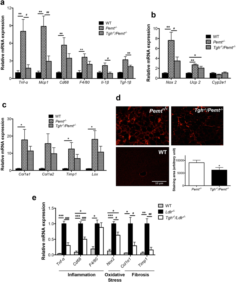
Figure 6. TGH deficiency ameliorates NASH features in Pemt−/− mice and Ldlr−/− mice.
(a) qPCR analysis of inflammatory genes expression in livers of WT, Pemt−/− and Tgh−/−/Pemt−/− mice after 10 weeks of HFD feeding, (b) qPCR analysis of oxidative stress genes expression in livers of WT, Pemt−/− and Tgh−/−/Pemt−/− mice, (c) qPCR analysis of fibrosis genes expression in livers of WT, Pemt−/− and Tgh−/−/Pemt−/− mice. n = 5. Data are mean ± SEM. *P < 0.05, **P < 0.01 vs WT control, #P < 0.05, ##P < 0.01 vs Pemt−/− mice. (d) Representative picrosirius red (PSR)-stained liver sections, and averaged collagen volume fraction in Pemt−/− and Tgh−/−/Pemt−/− mice. n = 5. Data are mean ± SEM. *P < 0.05, vs Pemt−/− mice. (e) qPCR analysis of representative NASH genes in livers of Ldlr−/− and Tgh−/−/Ldlr−/− after 12 weeks of WTD. n = 5. Data are mean ± SEM. *P < 0.05, **P < 0.01, ***P < 0.001 vs Ldlr−/− mice.

