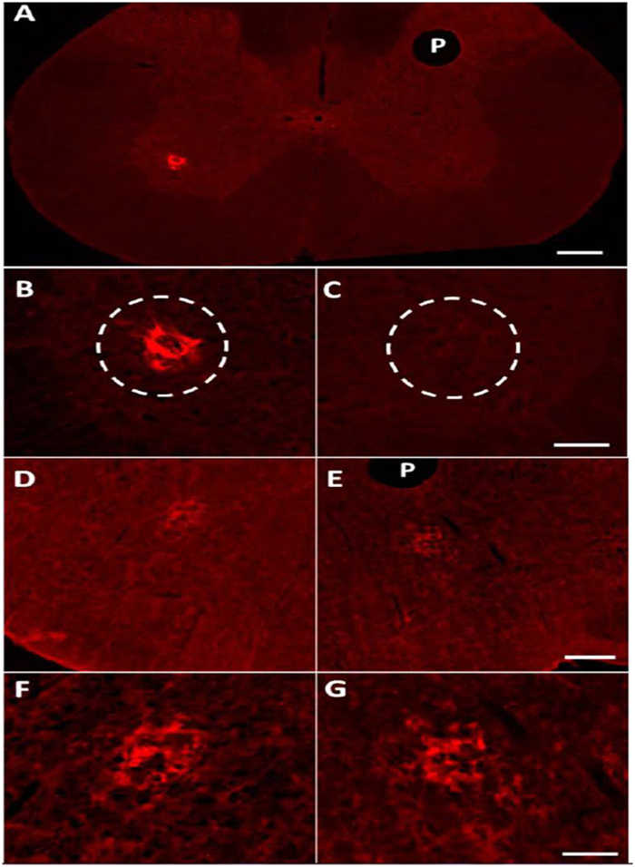
Figure 4.
(A) Transverse section of the cervical spinal cord displaying ipsilateral anti-WGA positive labeling in the phrenic nuclei (PN) following intradiaphragmatic injection of the WGA-HRP-AuNP-pro-THP nanoconjugate. There is a lack of labeling in the contralateral PN. Scale bar is 200 μm, P notes pinhole to mark side contralateral to the injection. (B) Higher magnification of the ipsilateral PN shown in A displaying fluorescence from the anti-WGA antibody. (C) Higher magnification of the contralateral PN shown in (A)with a complete lack of WGA label. Scale bar = 50 μm. (D,E) Transverse sections of the medulla at the level of the rVRGs from the same rat shown in A. (D) Ipsilateral rVRG displaying fluorescence from the anti-WGA antibody. (E) Contralateral rVRG displaying fluorescence from the WGA antibody. P notes pinhole to mark side contralateral to the injection. Scale bar = 200 μm. (F) Higher magnification of the ipsilateral rVRG show in (D). (G) Higher magnification of the contralateral rVRG shown in E. Scale bar = 50 μm.

