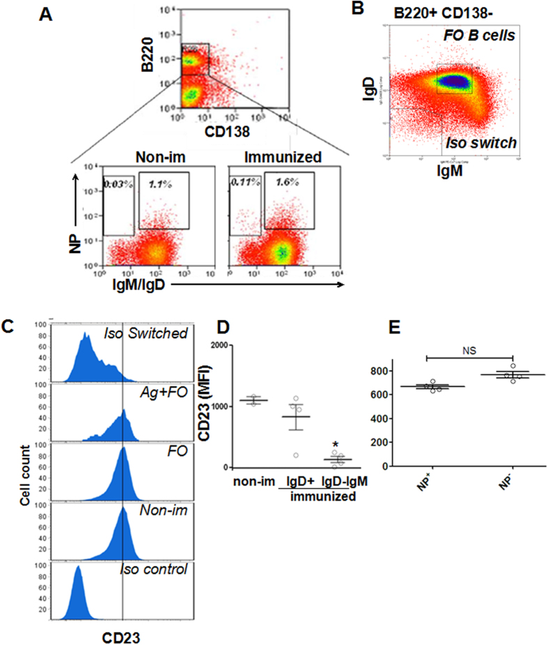
Figure 1. The expression levels of CD23 are reduced in isotype switched and memory B cells compared to follicular unswitched B cells.
The expression levels of CD23 in indicated B-cell subsets from a representative mouse 100 days post first immunization were determined using flow cytometry. Splenic B cells were isolated from non-immunized mice and mice 100 days post the first immunization with NP-KLH and Ribi adjuvant. Cells were stained with FITC-anti-IgD, FITC-anti-IgM, PE-anti-CD138, PerCP-Cy5.5-anti-B220 Abs and allophycocyanin-NP19 (A). B-cell subsets were identified by specific surface markers: NP-binding memory B cells (Memory) B220+ CD138− IgM− IgD− NP+, NP-binding unswitched follicular B cells (Ag+ FO) B220+ CD138− IgMint IgD+ NP+, naïve follicular B cells (FO) B220+ CD138− IgMint IgD+ NP− (A,B). Shown are representative histograms (C), the dot blot to show the gating stretagy(A and B), and the average (±SD) of the mean fluorescence intensity (MFI) of CD23 in follicular B cells from non-immunized mice (non-im), and follicular isotype unswitched (IgD+) and switched (IgD−IgM−) B cells (D), and NP+ or NP− B cells (E) from immunized mice. n = 4. **p < 0.01.

