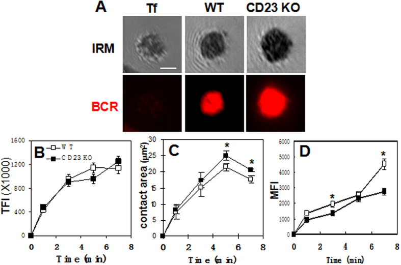
Figure 2. Effects of CD23 gene deletion on B-cell morphology and BCR clustering.
Splenic B cells from WT and CD23 KO mice were incubated with AF546-mB-Fab’-anti-Ig tethered lipid bilayers at 37 °C. As a non-antigen control, WT B cells were labeled with AF546-Fab’-anti-Ig for the BCR before incubation with biotinylated transferrin (Tf) tethered to lipid bilayers. The B-cell contact area and the fluorescence intensity of AF546-mB-Fab’-anti-Ig in the contact zone were quantified. Shown are representative images of cells at 7 min (A) and the average values (±SD) of the total (TFI) (B) and the contact area (C) and mean fluorescence intensity (MFI) of AF546-mB-Fab’-anti-Ig in the contact zone (D) from 50 cells of three independent experiments. Scale bar, 2.5 μm. *p < 0.05.

