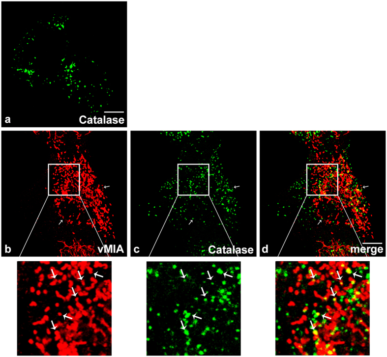
Figure 3. vMIA localization upon HCMV infection.
(a) Representative image of peroxisomal morphology in uninfected HFF cells, stained with catalase. (b–d) HFF cells infected with HCMV, 8 h post-infection. (b) vMIA, (c) catalase and (d) merge image of b and c. Arrows indicate co-localization loci. Bar represents 10 μm.

