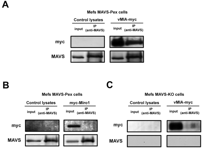
Figure 6. Interactions between peroxisomal MAVS and vMIA.
(A) Co-immunoprecipitation analysis of the interaction between overexpressed vMIA-myc and endogenous MAVS in Mefs MAVS-Pex cells. Negative control was performed by immunoprecipitating non-transfected cells. The pull-down was performed using an antibody against MAVS. Western blot was performed with antibodies against MAVS and myc. Input represents total cell lysate and IP represents the immunoprecipitation. (B) As negative control, the mitochondrial myc-tagged Miro1 (myc-Miro1) was transfected in Mefs MAVS-Pex cells. The pull-down was performed using an antibody against MAVS. Western Blot was performed with antibodies against MAVS and myc. Input represents total cell lysate and IP represents the immunoprecipitation. (C) As negative control, vMIA-myc was transfected in Mefs MAVS-KO cells. The pull-down was performed using an antibody against MAVS. Western Blot was performed with antibodies against MAVS and myc. Input represents total cell lysate and IP represents the immunoprecipitation.

