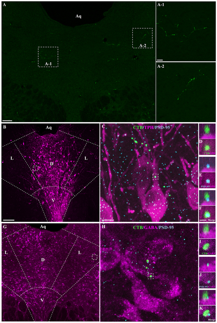
Figure 1. DRN-projecting RGCs contact 5-HT and GABA neurons in the DRND/L.
(A) CTB-labeled RGC processes in DRN. (B) Z-stack image of TPH neurons (magenta) in DRND/L. Dorsal raphe nucleus, lateral: L; dorsal raphe nucleus, dorsal: D; dorsal raphe nucleus, ventral: V. (C) Box in (B). Note a CTB-labeled RGC process associated with TPH neurons (Green: CTB; Blue: PSD-95). (D–F) Box in C. (D–F) Note that a CTB+ bouton is in apposition to a PSD-95 puncta on a TPH+ neuron. (G) Z-stack image of GABA neurons (magenta) in DRND/L. (H) Box in (G). Note a CTB-labeled RGC process associated with GABA neurons (Green: CTB; Blue: PSD-95). (I–K) Box in H. (I–K) Note that a CTB+ bouton is in apposition to PSD-95 puncta on a GABA+ neuron. Aq: aqueduct. Scale bars: 100 μm in A; 20 μm in A-1 (applies to A-2); 100 μm in B (applies to G); 2 μm in C (applies to H); 1 μm in F (applies to D, E and I to K).

