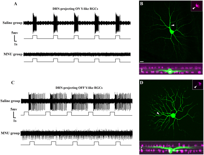
Figure 3. Effect of MNU on the activity of DRN-projecting RGCs.
(A) Firing patterns of DRN-projecting ON Y-like RGCs in response to visual stimulation. Top trace: Transient discharge pattern of retrogradely labeled ON Y-like RGC (firing increases at light onset) in a saline treated animal. Bottom trace: Loss of light-evoked firing of a retrogradely labeled ON Y-like RGC in an MNU-treated animal (representative of 37 recorded ON cells. (B) Dendritic morphology of the recorded DRN-projecting ON Y-like RGC in bottom trace. Arrow in inset at upper right corner points to the CTB retrogradely labeled soma. White dotted lines in lower plane indicated borders of sublamina a and b of IPL. Cholinergic amacrine cells were labeled and colored magenta. (C) Firing patterns of DRN-projecting OFF Y-like RGCs in response to visual stimulation. Top trace: Transient discharge pattern of retrogradely labeled OFF Y-like RGC (firing increases at light offset) in a saline treated animal. Bottom trace: Loss of visual responsiveness and vigorous spontaneous firing of a retorgradely labeled OFF Y-like RGC in an MNU treated animal (representative of 10 recorded OFF cells). (D) Dendritic morphology of the recorded DRN-projecting OFF Y-like RGC. Arrow in inset at upper right corner points to the CTB retrogradely labeled soma. White dotted lines in lower plane indicated borders of sublamina a and b of IPL. Cholinergic amacrine cells were labeled and colored magenta. Scale bar: 20 μm in B, applies to D.

