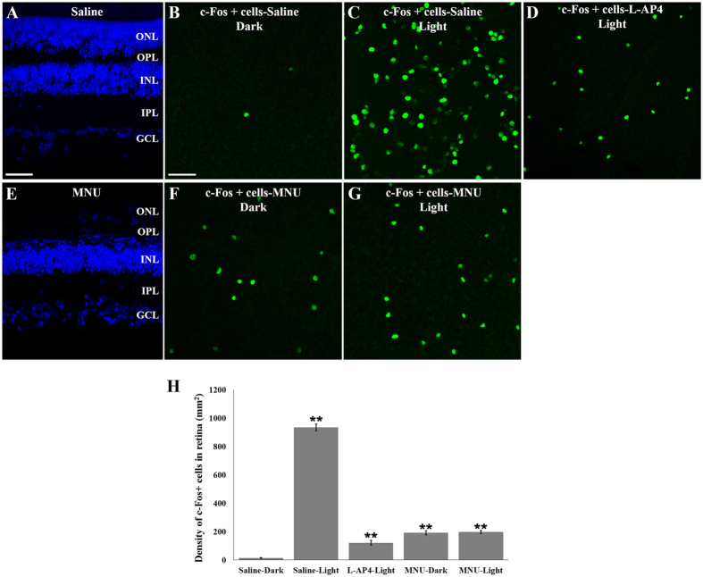
Figure 5. Effect of MNU on outer retina and c-Fos expression in ganglion cell layer.
(A) Normal retina of Saline-treated animal and (E) retina lacking outer nuclear layer (ONL) in MNU-treated animal. (B–D) c-Fos+ cells in the ganglion cell layer (GCL) of Saline-treated animals in the dark (B), after light stimulation (C) and in L-AP4 treated animals after light stimulation (D). (F–G) c-Fos+ cells in the GCL of MNU-treated animals in the dark (F) and after light stimulation (G). (H) Quantification of c-Fos+ cells; n = 5 retinas/group; (*p < 0.05; **p < 0.001) vs Saline-Dark group. Scale bars: 50 μm in A (applies to E); 20 μm in B (applies to C, D, F and G).

