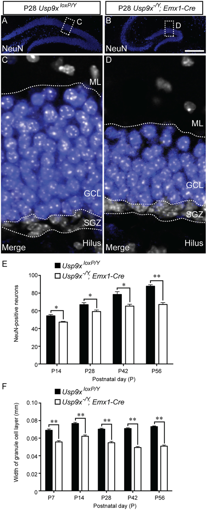
Figure 7. Fewer NeuN-expressing neurons in the dentate gyrus of Usp9x−/Y; Emx1-Cre mice.

(A,B) Mature neurons within the dentate gyrus of P28 Usp9xloxP/Y (A) and Usp9x−/Y; Emx1-Cre (B) are shown via NeuN labeling (blue). (C,D) Higher magnification views of the boxed regions in (A,B) respectively. These panels show the expression of NeuN (blue) and DAPI (white), revealing that the granule cell layer (GCL) is markedly thinner in mutant mice in comparison to controls. (E) Quantification of the number of NeuN-expressing neurons within the dentate gyrus revealed that there were significantly fewer neurons in the mutant at each of the ages studied. (F) Analysis of the width of the granule cell layer of the dentate gyrus revealed that this was significantly reduced in the mutant in comparison to controls at each of the ages assessed. *p < 0.05; **p < 0.01, t-test. SGZ – subgranular zone; ML – molecular layer. Scale bar in (B) (A,B) −150 μm; (C,D) −25 μm.
